Abstract
Techniques for controlling the rate and duration of drug delivery, while targeting specific locations of the body for treatment, to deliver the cargo (drugs or DNA) to particular parts of the body by what are becoming called “smart drug carriers” have gained increased attention during recent years. Using such smart carriers, researchers have also been investigating a number of physical energy forces including: magnetic fields, ultrasound, electric fields, temperature gradients, photoactivation or photorelease mechanisms, and mechanical forces to enhance drug delivery within the targeted cells or tissues and also to activate the drugs using a similar or a different type of external trigger. This review aims to cover a number of such physical energy modalities. Various advanced techniques such as magnetoporation, electroporation, iontophoresis, sonoporation/mechnoporation, phonophoresis, optoporation and thermoporation will be covered in the review. Special emphasis will be placed on photodynamic therapy owing to the experience of the authors’ laboratory in this area, but other types of drug cargo and DNA vectors will also be covered. Photothermal therapy and theranostics will also be discussed.
Keywords: Electroporation; Gene transfection; Magnetoporation; Nanoparticles; Optoporation; Photothermal therapy; Smart drug carriers; Sonoporation; Thermoporation.
Figures
Figure 1.

Different physical energy modules used for drug delivery
Drug delivery enhancers such as electroporation, magnetoporation, thermoporation, sonoporation and optoporation are illustrated.
Figure 2.
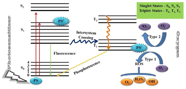
Jablonski Diagram
In the presence of molecular ground (triplet) state oxygen (3O2), the excited state PS transfers energy or electrons to produce reactive oxygen species (ROS).
Figure 3.
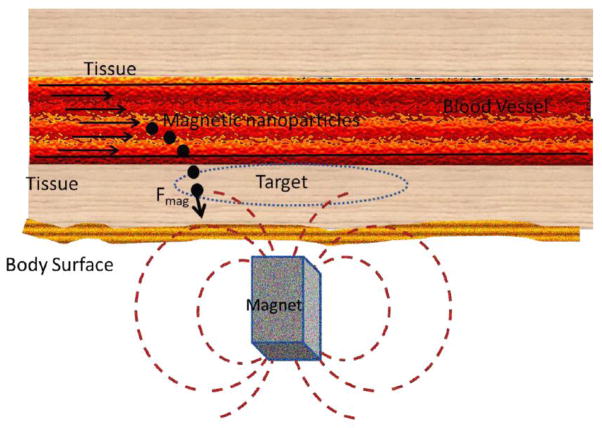
Schematic representation of a magnetic nanoparticle-based drug delivery system
Steps for magnetic nanoparticle-based drug delivery are; coupling the drug to the magnetic NP (MNP), applying an external magnetic field to attract the MNP to the desired location such as a tumor, and the release of the drugs from the NP under the influence of external alternating magnetic field once it reaches the target.
Figure 4.
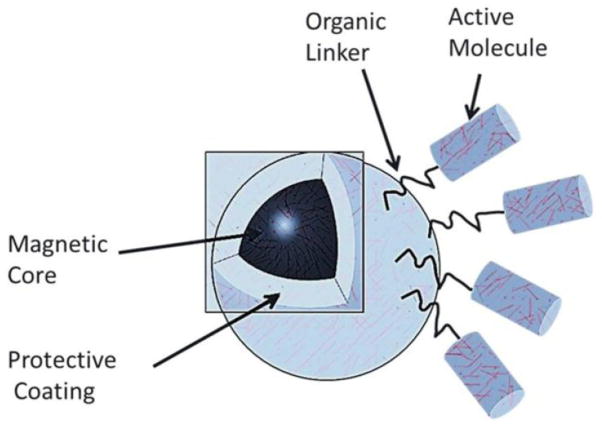
A typical magnetic nanoparticle
Magnetic nanoparticle consists of a magnetic core, a protective coating, an organic linker and an active molecule.
Figure 5.
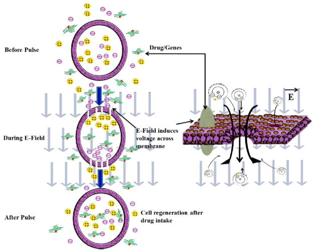
Electroporation phenomenon
Electric field disrupts cell membrane allowing drug/genes poration. The status of drug/genes before pulse, during the electric field (E-field) and after pulse is shown.
Figure 6.
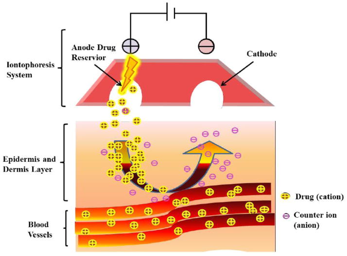
Iontophoresis method
The iontophoresis unit carries an anode drug reservoir that holds and releases the drug and a cathode that collects the opposite ions when a potential is applied across the two polarities.
Figure 7.
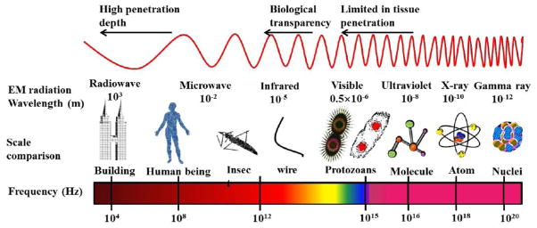
Light penetration as a function of wavelength
There exists a ‘window of transparency’ for human tissues in the far-red spectrum light is still limited in its penetration depth, or how far in can penetrate into the human body.
Figure 8.
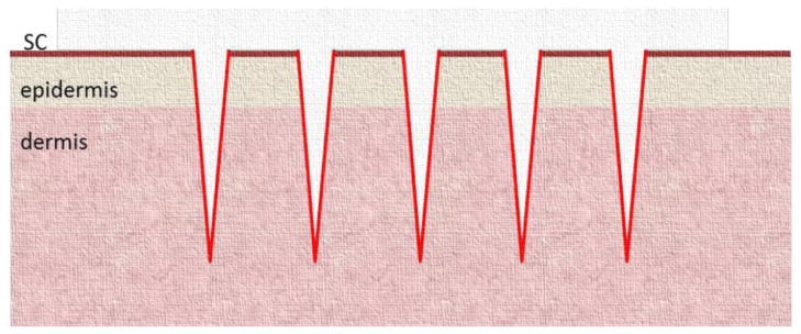
Optoporation
Schematic representation of micro-channels created by ablative fractional lasers in skin.
The light beam disrupts the major skin barrier, the stratum corneum (SC), and reaches deeper layers (dermis).
Figure 9.
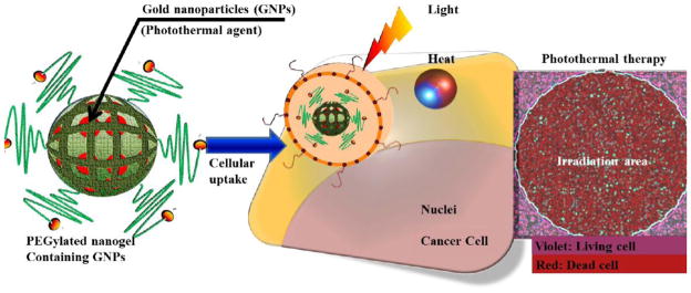
Photothermal therapy
PEGylated nanogels containing gold nanoparticels are uptaken by the cells. Irradiation (both heat and light) kills the cancer cells.
Figure 10.
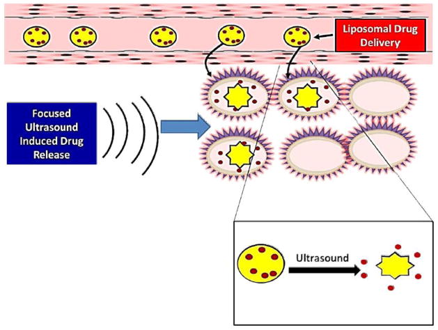
Sonoporation
Liposome bound drug is delivered to desired site. Ultrasound-sensitive liposomes burst open upon exposure to ultrasound leading to effective local delivery of the drug.
Figure 11.
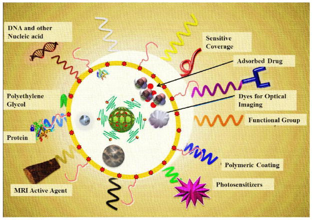
Nanoplatforms with theranostic functionalities
Fe2O3, magnetic NP, photosensitizer, appropriate polymeric coating, surface-charge tuning groups, optical imaging dyes, targeting agents, adsorbed/bonded drugs, sensitive coverage, DNA and other nucleic acids, polyethylene glycol, proteins and MRI active agents.